 DeepBio得分:5567.2
 DeepBio得分
是基于文献引用次数,影响因子,文献新近度等因素计算的客观产品评级,得分越高表明该产品经过越可靠的实验验证,质量可信度越高
DeepBio得分
是基于文献引用次数,影响因子,文献新近度等因素计算的客观产品评级,得分越高表明该产品经过越可靠的实验验证,质量可信度越高
产品属性
|
退换货说明 : 不支持退换货 |
性状 : 固体 |
存储条件 : ﹣20° |
|
保质期 : 1年 |
GFP antibody is reactive against all variants of Aequorea victoria GFP such as S65T-GFP, RS-GFP, YFP, CFP, RFP and EGFP.
Recombinant full length protein corresponding to GFP. Green fluorescent protein (GFP) from Aequorea victoria.
Database link: P42212
The total IgG concentration has been determined to be 5 mg/mL. The specific IgG concentration is unknown. This product should be kept refrigerated at all times whilst in short term storage. Using sterilised equipment will reduce the risk of bacterial contamination.
| 应用 | Ab评论 | 说明 |
|---|---|---|
| ICC/IF | (32) |
1/200 - 1/1000.
We recommend Goat Anti-Rabbit IgG H&L (Alexa Fluor® 488) (ab150081) secondary antibody. |
| ELISA |
Use at an assay dependent concentration.
|
|
| IHC-Fr | (8) |
Use at an assay dependent concentration.
Reported to work at dilutions up to 1/3000. Use secondary antibody Goat Anti-Rabbit IgG H&L (Alexa Fluor® 488) (ab15077). |
| IHC-P | (24) |
1/500 - 1/1000. Perform heat mediated antigen retrieval via the microwave method before commencing with IHC staining protocol.
|
| IP | (24) |
Use at an assay dependent concentration. Use at 1µl per 10cm tissue culture dish (use 10µl protein A agarose CL4B to precipitate the immune complex).
|
| WB | (70) |
1/1000 - 1/2500.
It is recommended to use 12.5% SDS-PAGE and to transfer to PVDF membrane. Use 1x Blotto (or 3% BSA in PBS) for diluting and blocking. Use PBS in 3x 5min washing steps throughout the immunolabelling. Probe with ab290 at 1:1000 - 1:5000 dilution and use Goat Anti-Rabbit IgG H&L (HRP) (ab205718) at 1:5000 dilution with ECL detection method. ab290 has been reported to work at 1:50,000 and dilutions around this range should be tested if high background is seen. Both incubation steps should be for 1hr at 22°C. |
| IHC-FoFr | (5) |
1/200 - 1/500.
|
| IHC-FrFl | (2) |
Use at an assay dependent concentration.
|
| Electron Microscopy |
1/1000 - 1/4000.
|
| 说明 |
|---|
|
ICC/IF
1/200 - 1/1000. We recommend Goat Anti-Rabbit IgG H&L (Alexa Fluor® 488) (ab150081) secondary antibody. |
|
ELISA
Use at an assay dependent concentration. |
|
IHC-Fr
Use at an assay dependent concentration. Reported to work at dilutions up to 1/3000. Use secondary antibody Goat Anti-Rabbit IgG H&L (Alexa Fluor® 488) (ab15077). |
|
IHC-P
1/500 - 1/1000. Perform heat mediated antigen retrieval via the microwave method before commencing with IHC staining protocol. |
|
IP
Use at an assay dependent concentration. Use at 1µl per 10cm tissue culture dish (use 10µl protein A agarose CL4B to precipitate the immune complex). |
|
WB
1/1000 - 1/2500. It is recommended to use 12.5% SDS-PAGE and to transfer to PVDF membrane. Use 1x Blotto (or 3% BSA in PBS) for diluting and blocking. Use PBS in 3x 5min washing steps throughout the immunolabelling. Probe with ab290 at 1:1000 - 1:5000 dilution and use Goat Anti-Rabbit IgG H&L (HRP) (ab205718) at 1:5000 dilution with ECL detection method. ab290 has been reported to work at 1:50,000 and dilutions around this range should be tested if high background is seen. Both incubation steps should be for 1hr at 22°C. |
|
IHC-FoFr
1/200 - 1/500. |
|
IHC-FrFl
Use at an assay dependent concentration. |
|
Electron Microscopy
1/1000 - 1/4000. |


Bone marrow-derived infiltrating cells in the stromal tissue of gastric intraepithelial tumor traced by GFP direct fluorescence.
(A) Normal tissues of the glandular stomach of a regular GFP(−) control mouse. (B) Normal tissues of the glandular stomach of a GFP(+) transgenic control mouse; (C, E, D, F) An induced gastric intraepithelial neoplasia (GIN) in a bone marrow transplanted mouse. GFP(+) BMDCs tracked with direct fluorescence localized in the GIN stromal tissue are shown in C and E. The same GIN lesion slide stained by H&E after the fluorescence observation are shown in D and F. DAPI (A–C and E) and hematoxylin (D and F) are used to visualize nuclei, respectively. Locations of the images C and D in the images E and F, and the image E in the image F are marked in the corresponding color. The gastric glands and stromal cells are also labeled.

GFP Immunohistochemistry (Free Floating) analysis of mouse brain tissue sections with ab290. Tissue was fixed with 4% PFA, frozen 30 µm sections were blocked for 1 hour at room temperature with 10% normal goat serum + donkey anti-mouse IgG Fab fragments (0.1 mg/ml). Sections were incubated with the primary antibody at a dilution of 1/1000 in TBS + 0.25% Triton-X for 16 hours at 4°C. A Cy2®-conjugated donkey anti-rabbit IgG (H+L) at a dilution of 1/200 was used as the secondary antibody.
Image shows anti-NeuN (red), DAPI (blue), and anti-GFP staining of GFP-cre (green, yellow with NeuN colocalization).

GFP staining with ab290 in dog hearts (Adv-GFP injection) tissue sections by IHC-P. Sections were PFA fixed and subjected to heat mediated antigen retrieval in citric acid (Ph6.0, 0.05% Tween20) prior to blocking with 10% serum for 30 mins at 37°C. The primary antibody was diluted 1/1000 in PBS and incubated with the sample for 1 hour at 25°C. A HRP conjugated secondary like Goat Anti-Rabbit IgG H&L (HRP) (ab205718) was used.

GFP immunofluorescence with GFP antibody ab290, images showing similar localization of Yes-GFP (first 10 aa's of Yes PTK fused to the N-terminus of GFP) to full length Yes PTK. A: Distribution of Yes detected using mouse anti-Yes Ab followed by Texas Red-conjugated anti-mouse Ab. B: Chimeric GFP's detected using rabbit anti-GFP Ab (Abcam ab290) followed by FITC-conjugated anti-rabbit Ab.
Image kindly provided by L.G. Berthiaume. Taken from J. McCabe and L.G. Berthiaume, Functional Roles for Fatty Acylated Amino-terminal Domains in Subcellular Localization, Molecular Biology of the Cell 10:3771-3786, 1999

GFP immunoprecipitation with ab290 in HEK293 nuclear lysate expressing GFP. 20µg of lysate was incubated with primary antibody (1 µg/mg lysate) and matrix (Protein G) for 16 hours at 4°C in AFC low salt buffer. For western blotting ab290 (1/5000) was used to confirm successful immunoprecipation.
Lane 1: HEK293 nuclear lysate expressing GFP input.
Lane 2: IP of HEK293 nuclear lysate expressing GFP.
Lane 3: Cells with no GFP.

GFP staining with GFP antibody ab290 in GFP-transfected NIH3T3 cells. The cells were fixed with 4% formaldehyde (10min) and then blocked in 1% BSA / 0.3M glycine in 0.1%PBS-Tween for 1h. The cells were then incubated with ab290 at 1/200 dilution overnight at +4°C followed by incubation with Goat Anti-Rabbit IgG H&L (Alexa Fluor® 488) (ab150081), for 1 hour, at 1μg/ml.
Under identical experimental conditions, when compared to the basal level of GFP expression in transfected NIH3T3 cells, the cells upon which ab290 was applied gave a stronger signal in the 488 channel, indicating that ab290 is binding to GFP and therefore eliciting signal amplification.
ab290 was also applied to non-GFP-transfected NIH3T3 cells, which produced no positive staining, indicating specificity for GFP. Nuclear DNA was labelled with 1.43μM DAPI (blue).

GFP immunoprecipitation with GFP antibody ab290 in human HEK293 cells transfected with Annexin1-GFP. 25µg of cell lysate was incubated with the primary antibody and matrix (Protein G) in 1% TX-100, 10% glycerol, 1X PBS for 16 hours at 4°C. For Western blotting anti-rabbit HRP conjugated secondary antibody was used at a dilution at 1/5000.
Lane 1: Lysate of HEK293 cells expressing Annexin1-GFP fusion protein.
Lane 2: IP with anti-GFP.
Lane 3: Not bound fraction.
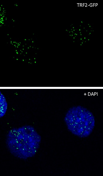
GFP staining with GFP antibody ab290 in U2OS cells expressing TRF2-GFP fusion protein by ICC/IF (Immunocytochemistry/immunofluorescence). Cells were fixed with formaldehyde, permeabilized with NP40 and blocked with 3% BSA for 1 hour at 21°C. Samples were incubated with the primary antibody (1/1000 in PBS + 3% BSA) for 12 hours at 4°C. An Goat Anti-Rabbit IgG H&L (Alexa Fluor® 488) (ab150077) at a dilution of 1/500 was used as the secondary antibody.
Green - GFP.
Blue - DAPI.
| 好评度 | 商品满意度 | 服务满意度 | 发货满意度 |

"Anti-GFP antibody"商品可能已被商家删除,您可查看其他相似商品!
相似产品推荐
 分类纠错
分类纠错

