 DeepBio得分:5567.2
 DeepBio得分
是基于文献引用次数,影响因子,文献新近度等因素计算的客观产品评级,得分越高表明该产品经过越可靠的实验验证,质量可信度越高
DeepBio得分
是基于文献引用次数,影响因子,文献新近度等因素计算的客观产品评级,得分越高表明该产品经过越可靠的实验验证,质量可信度越高
产品属性
|
退换货说明 : 不支持退换货 |
性状 : 固体 |
存储条件 : ﹣20° |
|
保质期 : 1年 |
From Jan 2024, QC testing of replenishment batches of this polyclonal changed. All tested and expected application and reactive species combinations are still covered by our Abcam product promise. However, we no longer test all applications. For more information on a specific batch, please contact our Scientific Support who will be happy to help.
Synthetic peptide. This information is proprietary to Abcam and/or its suppliers.
(Peptide available as
Ab15580 is batch tested in ICC/IHC. A variability in IHC-Fr performance can occur with this antibody but we can guarantee consistency in IHC-P. Some customers have successfully used ab15580 in Western Blot. Higher molecular weight proteins like Ki67 may be more difficult to detect in WB. We recommend several potential optimisation steps: loading higher amounts of protein (20 µg and above), using lower percentage gels and/or Tris-Acetate gels, increasing antioxidant to maintain protein reduction, decreasing methanol and increasing SDS in the transfer buffer, and increasing time and voltage of transfer. Larger proteins can be subject to degradation more than smaller proteins so lower molecular weight bands may be present. For further information or support please contact our Scientific Support Team.
The Life Science industry has been in the grips of a reproducibility crisis for a number of years. Abcam is leading the way in addressing this with our range of recombinant monoclonal antibodies and knockout edited cell lines for gold-standard validation. Please check that this product meets your needs before purchasing.
If you have any questions, special requirements or concerns, please send us an inquiry and/or contact our Support team ahead of purchase. Recommended alternatives for this product can be found below, along with publications, customer reviews and Q&As
Abpromise™承诺保证使用ab15580于以下的经测试应用
“应用说明”部分 下显示的仅为推荐的起始稀释度;实际最佳的稀释度/浓度应由使用者检定。
| 应用 | Ab评论 | 说明 |
|---|---|---|
| IHC-P | (68) |
Use a concentration of 0.1 - 5 µg/ml. Perform heat mediated antigen retrieval before commencing with IHC staining protocol.
|
| ICC/IF | (8) |
Use a concentration of 0.5 - 1 µg/ml.
If fixing cells in 4% PFA (20 min, room temp), it is recommended to permeabilized cells with 0.1% Triton-X for 5 min. Positive Control: HeLa and HAP1 cells |
| 说明 |
|---|
|
IHC-P
Use a concentration of 0.1 - 5 µg/ml. Perform heat mediated antigen retrieval before commencing with IHC staining protocol. |
|
ICC/IF
Use a concentration of 0.5 - 1 µg/ml. If fixing cells in 4% PFA (20 min, room temp), it is recommended to permeabilized cells with 0.1% Triton-X for 5 min. Positive Control: HeLa and HAP1 cells |
Target information above from: UniProt accession
P46013
The UniProt Consortium
The Universal Protein Resource (UniProt) in 2010
Nucleic Acids Res. 38:D142-D148 (2010)
.


ab15580 staining Ki67 in Mef1 cells. The cells were fixed with 100% methanol (5 min), permeabilized with 0.1% PBS-Triton X-100 for 5 minutes and then blocked with 1% BSA/10% normal goat serum/0.3M glycine in 0.1%PBS-Tween for 1h. The cells were then incubated overnight at 4°C with ab15580 at 0.5 µg/ml and ab7291, Mouse monoclonal [DM1A] to alpha Tubulin - Loading Control. Cells were then incubated with ab150081, Goat polyclonal Secondary Antibody to Rabbit IgG - H&L (Alexa Fluor® 488), pre-adsorbed at 1/1000 dilution (shown in green) and ab150120, Goat polyclonal Secondary Antibody to Mouse IgG - H&L (Alexa Fluor® 594), pre-adsorbed at 1/1000 dilution (shown in pseudocolour red). Nuclear DNA was labelled with DAPI (shown in blue).
Also suitable in cells fixed with 4% paraformaldehyde (10 min).
Image was acquired with a high-content analyser (Operetta CLS, Perkin Elmer) and a single confocal section is shown.

ab15580 staining Ki67 in HeLa cells. The cells were fixed with 100% methanol (5 min), permeabilized with 0.1% PBS-Triton X-100 for 5 minutes and then blocked with 1% BSA/10% normal goat serum/0.3M glycine in 0.1%PBS-Tween for 1h. The cells were then incubated overnight at 4°C with ab15580 at 0.5 µg/ml and ab7291, Mouse monoclonal [DM1A] to alpha Tubulin - Loading Control. Cells were then incubated with ab150081, Goat polyclonal Secondary Antibody to Rabbit IgG - H&L (Alexa Fluor® 488), pre-adsorbed at 1/1000 dilution (shown in green) and ab150120, Goat polyclonal Secondary Antibody to Mouse IgG - H&L (Alexa Fluor® 594), pre-adsorbed at 1/1000 dilution (shown in pseudocolour red). Nuclear DNA was labelled with DAPI (shown in blue).
Also suitable in cells fixed with 4% paraformaldehyde (10 min).
Image was acquired with a high-content analyser (Operetta CLS, Perkin Elmer) and a maximum intensity projection of confocal sections is shown.

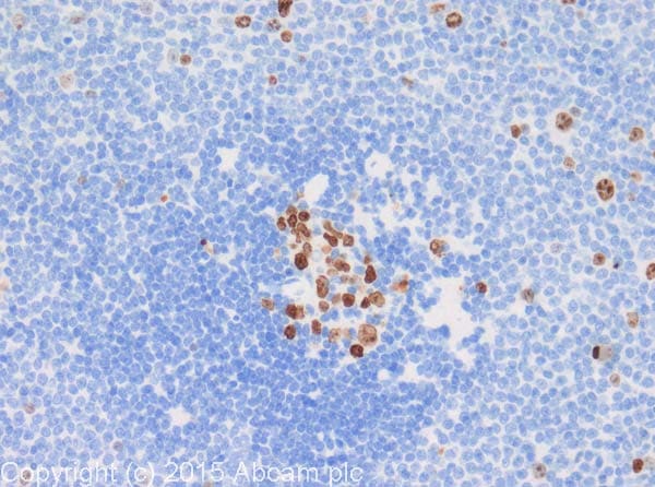
IHC image of ab15580 staining in mouse spleen formalin fixed paraffin embedded tissue section, performed on a Leica BondTM system using the standard protocol B. The section was pre-treated using heat mediated antigen retrieval with sodium citrate buffer (pH6) for 20 mins. The section was then incubated with ab15580, 5µg/ml, for 15 mins at room temperature. A goat anti-rabbit biotinylated secondary antibody was used to detect the primary, and visualized using an HRP conjugated ABC system. DAB was used as the chromogen. The section was then counterstained with haematoxylin and mounted with DPX.

Immunohistochemical analysis of formalin fixed paraffin embedded human tonsil labelling Ki67 with ab15580 at a concentration of 0.5 µg/ml. The immunostaining was performed on a Ventana DISCOVERY ULTRA (Roche Tissue Diagnostics) instrument with an OptiView DAB IHC Detection Kit. Heat mediated antigen retrieval was conducted for 32min with ULTRA cell conditioning solution (CC1 pH8.5). ab15580 anti Ki67 antibody was incubated at 37°C for 16min. Sections were counterstained is with Hematoxylin II. Image inset shows absence of staining in secondary antibody only control

Paraformaldehyde-fixed Rabbit cell (Retina) labeling Ki67 (Green) using ab15580 at 1/200 dilution followed by a Donkey anti-rabbit Alexa Fluor® 568 secondary antibody in ICC analysis. Normal Donkey serum was used as the blocking agent for 15 hours at 4ºC.
Tissue was immersion fixed in 4% paraformaldehyde overnight at 4 degrees Celsius. Tissue was then embedded in 10% agarose and section at 100 microns. Sections were placed in 2N HCL for 1 hour before commencing immunocytochemistry. Ki-67 (dividing cells red).
IHC image of Ki67 staining in human spleen formalin fixed paraffin embedded tissue section, performed on a Leica BondTM system using the standard protocol F. The section was pre-treated using heat mediated antigen retrieval with sodium citrate buffer (pH6) for 20 mins. The section was then incubated with ab15580, 1µg/ml, for 15 mins at room temperature and detected using an HRP conjugated compact polymer system. DAB was used as the chromogen. The section was then counterstained with haematoxylin and mounted with DPX.
For other IHC staining systems (automated and non-automated) customers should optimize variable parameters such as antigen retrieval conditions, primary antibody concentration and antibody incubation times.

ab15580 staining Ki67 in wild-type HAP1 cells (top panel) and Ki67 knockout HAP1 cells (bottom panel). The cells were fixed with 100% methanol (5min), permeabilized with 0.1% Triton X-100 for 5 minutes and then blocked with 1% BSA/10% normal goat serum/0.3M glycine in 0.1% PBS-Tween for 1h. The cells were then incubated with ab15580 at 1μg/ml concentration and ab195889 at 1/250 dilution (shown in pseudo colour red) overnight at +4°C, followed by a further incubation at room temperature for 1h with a goat anti-rabbit IgG Alexa Fluor® 488 (ab150081) at 2 μg/ml (shown in green). Nuclear DNA was labelled in blue with DAPI.

Confocal images of mouse trachea epithelium collected at stea
| 好评度 | 商品满意度 | 服务满意度 | 发货满意度 |

"Anti-Ki67 antibody"商品可能已被商家删除,您可查看其他相似商品!
相似产品推荐
 分类纠错
分类纠错

